Research Article - Modern Phytomorphology ( 2025) Volume 19, Issue 5
The gynoecium structure in Sansevieria grandicuspis Haw. (Asparagaceae)
Oksana Fishchuk* and N. KostrubaOksana Fishchuk, Lesia Ukrainka Volyn National University, Pr. Voli. 13, Lutsk, Ukraine, Email: Fishchuk.Oksana@vnu.edu.ua
Received: 17-Nov-2024, Manuscript No. mp-24-152748; , Pre QC No. mp-24-152748 (PQ); Editor assigned: 20-Nov-2025, Pre QC No. mp-24-152748 (PQ); Reviewed: 06-Dec-2025, QC No. mp-24-152748; Revised: 11-Sep-2025, Manuscript No. mp-24-152748 (R); Published: 15-Sep-2025, DOI: 10.5281/zenodo.17722285
Abstract
Gynoecium of Sansevieria grandicuspis and vertical zonality of the ovary, gynoecium structural zonality after W. Leinfellner and septal nectary zonality were studied. There are a high parenchymatous ovary base with septal nectaries, ovary locules-the biggest part of the ovary with three locule cavities and the cavity of septal nectary, ovary roof-the part over the locules. There are three style channels and the end of septal nectaries. There is a long septal nectary that can be prolongated in both of them and opens with the secretory nectary splits. The gynoecium of this species has a short synascidiate zone, fertile hemisynascidiate zone and asymplicate zone that embraces the ovary roof, common style and stigma. Since there is a debate about the belonging of the Sansevieria genus to the Dracaena genus according to molecular data, it is interesting to study the morphological and anatomical features of these genera for further comparison of the flower structure, namely the structure of the ovary and septal nectary structure and the confirmation or disconfirmation of the molecular data by anatomical and micro morphological data.
Keywords
Sansevieria, Gynoecium morphology, Gynoecium vertical zonality, Ovary structure, Septal nectary
Introduction
The study of representatives of the genus Sansevieria is very actual in the world, in particular, phytoremidation by three Sansevieria species through absorption of carbon monoxide (Iswoyo, et al. 2023), effect of alkali on the mechanical properties of Sansevieria fiber bio-composites and as a result it was proved that alkali treatment increased the potential of Sansevieria fibers as composite reinforcement (Widodo, et al. 2024). Scientists from Bangladesh studied biological model by four Sansevieria species (S. zeylanica, S. metallica, S. trifasciata S., roxburghiana) and considers that these plants are “natural herbal source” and they can be used as potential pharmacological applications in large-scale industries (Rajashekara, 2024), inhibition by Sansevieria trifasciata extract of a the growth of Escherichia coli bacteria, in vitro (Rachmaniyah and Rusmiati, 2023). An interesting study is the purification of the environment from PM 2.5 in a school by planting Sansevieria trifasciata (Sutrisno, et al. 2023).
Materials and Methods
Plant material Sansevieria grandicuspis was collected in the A.V. Fomin Botanical Garden of Taras Shevchenko National University of Kyiv and fixed in 70% alcohol. For light microscope observations 10 flowers S. grandicuspis were sectioned on transverse sections of 20 μm thickness were obtained with a manual rotary microtome and according to staining protocol (Soukup and Tylová, 2019) stained in Safranin and Astra Blue. Slides were mounted in “Eukitt®”. Images were obtained with an AMSCOPE 10 MP digital camera attached to an AMSCOPE T490B-10M (USA) microscope. Measurements of 10 fresh flowers were carried out for morphological analysis. We used the concept of gynoecium vertical zonality by Leinfellner, 1950 to analyze the gynoecium's internal structure. The height of the zones of gynoecium was measured according to the number of cross sections.
Results
Flowers of S. grandicuspis 11 mm-15 mm long, slightly zygomorphic. Bracts length of 2.5 mm, 1 mm wide, elongated, ovoid, leathery and light brown. Pedicel 0.75 mm-1 mm in diameter, 3.5 mm-4 mm in length, pedicel 2/3 length. Sepal one, rear, ovoid with a basis of 0.75 mm, a width is 1 mm, 1.25 long, light brown. Perianth is simple, 6-membered and white. Flower is like a tube (tubular), smaller mid increased, 1.3 mm-1.8 mm in diameter and 6 mm in length. Free parts of simple perianth is with curved tip, 1.5 mm-1.7 mm in width and 6,5-9 mm long. The stamens are separated from the floral tube slightly lower than stamen outer circle. The length of the free part of the external and internal stamens 4.5 mm-6.5 mm. Stamen filaments are 0.25 mm in diameter. Anthers are 1.6 mm-1.8 mm long, 0.25 mm wide. Filaments are attached to anther below the middle.
Gynoecium consists of three accrete carpels, each of them has one seed bud. Pistil is slightly zygomorphic. Ovary is superior and ovate, 1.5 mm-1.8 mm in diameter, 1.8 mm-2.2 mm long, wrinkled. The style is not in the central location, it is in the side of the ovary and wavy, up to 15 mm long, 0.3 mm-0.5 mm in diameter. Stigmas blades are linear and deflected, 0.25 mm long and 0.25 mm in width.
Gynoecium structure
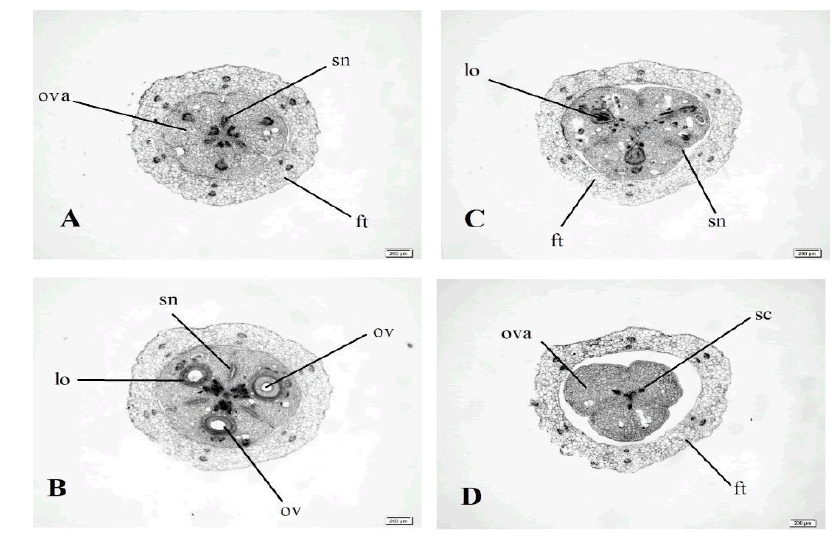
The gynoecium of S. grandicuspis is trimerous. Ovary is trilocular, with one median anatropous ovule in each locule and long septal nectary (Fig. 1). Each ovule has well developed funicular obturator (Fig. 1D). Three style channels go through the style (Fig. 1D). Stigma is trilobate, with close channels.
Figure 1: Ascending series of transversal sections of the flower S. grandicuspis. Scale bar 200 μm.
As in the previously studied species of Dracaena-Sansevieria group of Asparagaceae (Fishchuk and Odintsova, 2013), we have revealed three main parts of the ovary of the studied spesies: Ovary base (Fig. 1A) with septal nectaries, ovary locules– the biggest part of the ovary with three locule cavities and the cavity of septal nectary (Fig. 1B), ovary roof-the part over the locules. There are three style channels and the end of septal nectaries (Fig. 1D).
Accordingly, to the concept of vertical zonality of the gynoecium (Leinfellner, 1950), we researched three gynoecium vertical zones in S. grandicuspis:
Synascidiate zone: The short gynoecium zone (140 μm) with three disconnected locules. Here presented three septal nectary cavities (Fig.1A). As the locules, nectary cavities here have no common epidermises, and really in this zone there are six distinct cavities– three locules and three nectaries.
Hemisynascidiate zone: The longest gynoecium zone with three separated locules and a three-fold crack in the center (660 μm). In the central part there are two ranges of the epidermal cells. They are post genitally closed (Fig. 1B).
Asymplicate zone: Zone where carpels are melted only post genitally. The beginning of this zone is where each septal nectary cavity consolidates distally with the septal groove (Fig. 1C and 1D). This zone includes ovary roof, style and stigma and has no congenital fusion among carpels after (Leinfellner, 1950).
Discussion
The flowers in the genus Sansevieria had one ovule in each locule and three-dimensional structure of the floral nectary were presented (Odintsova, 2014). Sansevieria pfennigii, which was investigated by a group of scientists in the Lindi region in southern Tanzania and they made a detailed morphological description based on additional observations recorded from living plants, including fruits that were previously unknown (Scharf and Burkart, 2021).
In Sansevieria hyacinthoides gynoecium presence synascidiate, hemisynascidiate, hemisymplicate and asymplicate zones (Odintsova and Fishchuk, 2013). In Sansevieria suffruticosa gynoecium were found synascidiate zone, which was typical for the eusyncarpous gynoecium, hemisynascidiate (fertile), hemisymplicate and asymplicate zones (Fishchuk and Odintsova, 2013).
Studies of famous botanists presents the results of a micro morphological study on stamens of Sansevieria species. They described flowers of 15 species and proved that morphological features, in particular stamen micromorphology may be taxonomically significant (Klimko, et al. 2017). This group of scientists also described pollen grain morphology in 11 Sansevieria species for the first time in terms of pollen micromorphology (Klimko, et al. 2017).
Conclusion
In the S. grandicuspis ovary there were three structural ovary zones: A short synascidiate, fertile hemisynascidiate and asymplicate zone that embraces the ovary roof, common style and stigma Gynoecium consists of three accrete carpels, each of them has one seed bud. Pistil is slightly zygomorphic. Ovary is superior and ovate, wrinkled. The style is not in the central location, it is in the side of the ovary and wavy. Stigmas blades are linear and deflected. Our results confirm that floral morphology and anatomical features provides key taxonomic information to assess species delimitation in Dracaena and Sansevieria.
References
- Iswoyo H, Bahrun AH, Ganing W. (2023). Phytoremidation by Sansevieria sp. through absorption of Carbon Monoxide (CO). Agrovigor J Agroekoteknologi. 16:46-52.
- Widodo EW, Pratikto P, Sugiarto S, Widodo TD. (2024). Effect of alkali on the mechanical properties of Sansevieria fiber bio-composites. J Adv Res Appl Mech. 121:117-134.
- Rajashekara S. (2024). Sansevieria species (snake plants): An interesting biological model and a forthcoming good model genetic organisms for popularization, utilization and conservation. Environ Anal Ecol Stud. 12:1473-1477.
[Crossref]
- Rachmaniyah R, Rusmiati R. (2023). Sansevieria trifasciata extract effectively inhibit the growth of Escherichia coli bacteria, in vitro. Int J Public Health Sci. 12:1119-1124.
- Sutrisno S., Wiwaha G., Sofiatin Y. (2023). The effect of sansevieria plant on particulate matter 2.5 levels in classroom. J Kesehat Masy. 18:397-407.
- Soukup A, Tylová E. (2019). Essential methods of plant sample preparation for light microscopy. Methods Mol Biol. 1992:1-26.
- Leinfellner W. (1950). Der Bauplan des syncarpen Gynoeceums. Österreichische Botanische Zeitschrift. 97:403-436.
- Fishchuk O, Odintsova A. (2013). Morphology and vascular anatomy of the flower in Sansevieria suffruticosa N. E. Br. (Asparagaceae Juss.). Studia Biologica. 7:139-148.
- Odintsova A, Fishchuk O. (2013). Morphology and vascular anatomy of the flower in Sansevieria hyacinthoides (L.) Druce (Asparagaceae Juss.). Visnyk Lviv Univ. 62:99-107.
- Odintsova A, Fishchuk O, Sulborska A. (2014). Gynoecium structure in Dracaena fragrans (L.) Ker Gawl., Sansevieria parva N.E. Brown and Sansevieria trifasciata Prain (Asparagaceae) with septal emphasis on the structure of the septal nectary. Acta Agrobotanica. 66:55-64.
- Scharf U, Burkart M. (2021). Sansevieria pfennigii (Ruscaceae, Asparagales): Confirmation of existence, emendation of description, and tentative threat assessment. Phytotaxa. 483:1-8.
- Klimko M, Wysakowska I, Wilkin P, Wiland-SzymaÅska J. (2017). Micromorphology of stamens of some species of the genus Sansiviera petagna (Asparagaceae). Steciana. 21:41-48.
- Klimko M, Wysakowska I, Wilkin P, Wiland-SzymaÅska J. (2017). Pollen morphology of some species of the genus Sansevieria petagna (Asparagaceae). Acta Biol Cracs Bot. 59:63-75.
